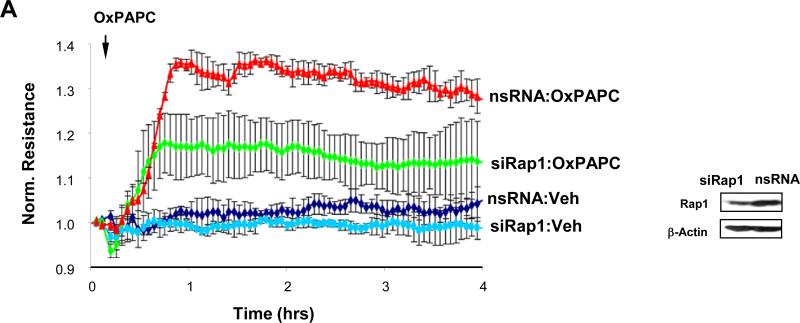
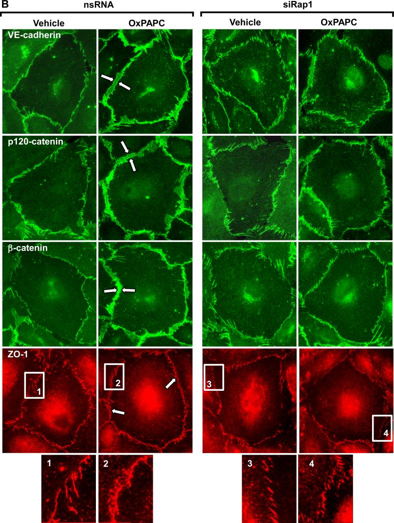
Figure 4. Effect of Rap1 depletion on OxPAPC-mediated adherens junction and tight junction remodeling.
Pulmonary EC were transfected with Rap1-specific siRNA, as described in Methods. Control cells were treated with non-specific RNA. A: HPAEC were stimulated with OxPAPC (10 μg/ml) or vehicle (indicated by arrow), and TER changes were monitored over 4 hrs. Inset depicts Rap1 protein depletion induced by specific siRNA duplexes, which was compared to treatment with non-specific RNA. Membrane reprobing with β-actin antibody was used as normalization control. Results are representative of five independent experiments. B: Cells were treated with OxPAPC for 30 min followed by immunofluorescence staining for VE-cadherin, p120-catenin, β-catenin, and ZO-1. High magnification insets depict ZO-1 staining of the cell-cell junctions in the nsRNA- and siRap1-treated cells. Shown are representative results of three independent experiments.


