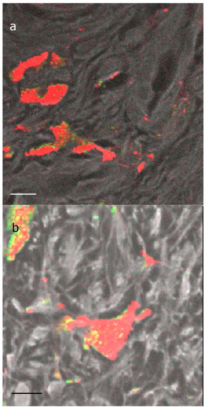
Figure 2. Macrophages containing melanin.
Images of macrophages containing melanin in a benign lesion (a) and a malignant melanoma (b). Color bar is the same as in figure 1. The black and white color scale, overlaid with the melanin channel in some cases, is the fluorescence and second harmonic generation channel, which serves as a structural reference for interpreting the images. Scale bar = 10 μm.

