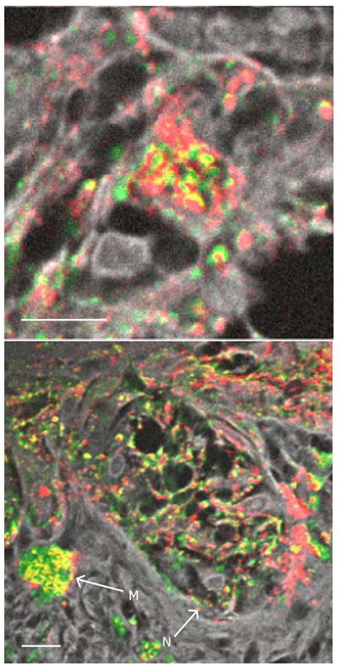
Figure 4. Melanin in a melanocytic nest.

(a) High magnification of a malignant melanocyte in a melanocytic nest; (b) malignant melanocytic nest with arrows marking a macrophage (M) and a melanocytic nest (N). Color scheme is the same as figure 2. Scale bar = 10 μm.
