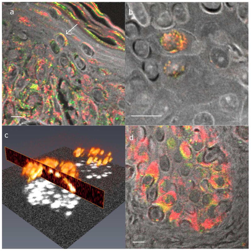
Figure 5. Examples of supranuclear melanin caps.

(a) Cross section of melanin caps on suprabasal cells. The arrow points to an example of a supranuclear cap. (b) En face supranuclear melanin cap. (c) Three-dimensional rendering of (b). (d) Cross section of melanin caps on basal cells. Images (a), (b), and (d) all have the same color scheme as figure 3, and the scale bars = 10 μm. (c) A rendering of the total melanin content, shown by the melanin signal at time zero.
