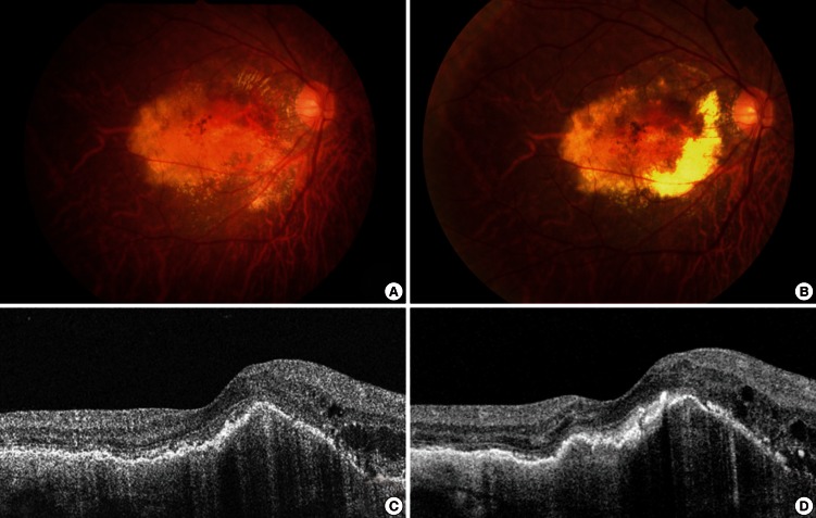
Fig. 1.
Fundus photographs and optical coherence tomography (OCT) images from a 57-yr-old male smoker. (A) A baseline fundus photograph shows submacular hemorrhage and exudate. (B) A post-treatment fundus photograph shows persistent submacular hemorrhage, extensive macular retinal pigment epithelial atrophy, and a disciform scar. Pre- (C) and post-treatment (D) OCT images show virtually no anatomical improvements following intravitreal ranibizumab treatment.

