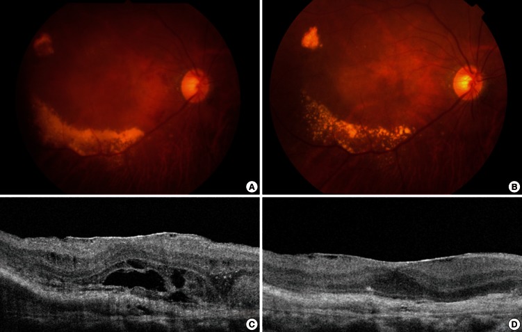
Fig. 2.
Fundus photographs and optical coherence tomography images (OCT) of a 73-yr-old male ex-smoker. (A) A baseline fundus photograph shows submacular hemorrhage, the choroidal neovascular membrane, and exudate. (B) A post-treatment fundus photograph shows resolution of the submacular hemorrhage, but choroidal neovascularization and exudate persists. (C) A baseline OCT image reveals subretinal fluid and a thickened choroidal neovascular membrane. (D) A post-treatment OCT image shows resolution of the previously observed subretinal fluid and thinning of a now inactive choroidal neovascular membrane.

