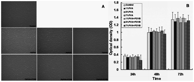
Figure 4.
(A) Cytotoxicity using L929 fibroblasts cell culture in different samples extracts (1% PVA, 3% PVA, 7% PVA, 1% PVA + FCVB, 3% PVA + FCVB, and 7% PVA + FCVB). No changes in cell morphology were observed in any groups (200×). (B) MTT assay at 24, 48 and 72 hours. No significant difference among sample extracts as compared to the negative control (P > 0.05) was observed.

