
Figure 2. Neuroimaging and neuropathologic findings in pigmented orthochromatic leukodystrophy.
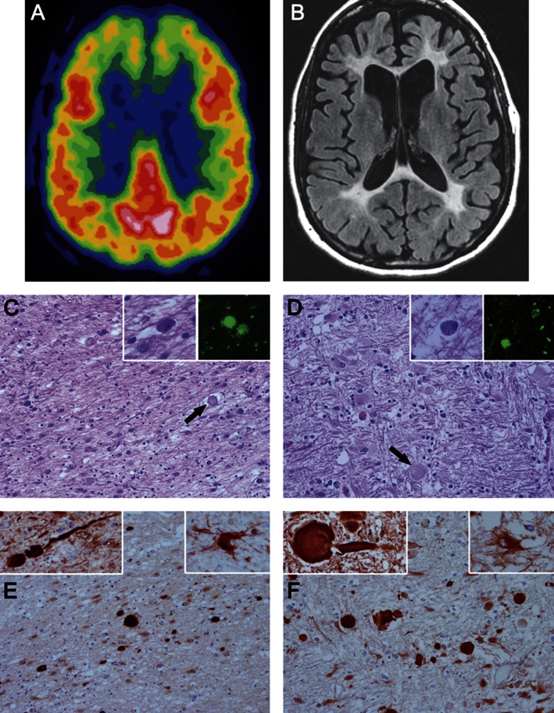
(A) 18-Fluorodeoxyglucose PET scan performed in 2006 from FTD368 II-1 shows hypometabolism in both frontal lobes. (B) MRI scan performed in 2007 shows atrophy of the frontal lobes, white matter hyperintensities in the white matter of the frontal lobes and similar changes in the parietooccipital white matter, enlargement of the frontal horns of the lateral ventricles, and milder diffuse cerebral atrophy. (C–F) Microscopic images of white matter from FTD368 II-2 (C and E) compared with typical hereditary diffuse leukoencephalopathy with axonal spheroids (HDLS) (D and F). On hematoxylin & eosin stains, the white matter has pallor because of myelinated fiber loss and gliosis, with axonal spheroids (arrows in C and D). Within the affected white matter are macrophages with basophilic to brown fine granules (left insets in C and D) that have autofluorescent pigment (right insets in C and D) when unstained sections are viewed with a fluorescent microscope. Immunohistochemistry for amyloid precursor protein (panels E and F) shows many axonal spheroids in affected white matter, which are confirmed to be axonal in origin by immunoreactivity with phosphorylated neurofilament (left insets in E and F). A characteristic feature of HDLS is the presence of bizarre hypertrophic astrocytes demonstrated with immunohistochemistry for αB-crystallin (right insets in E and F). C–F: original magnification ×200; all insets: original magnification ×400.