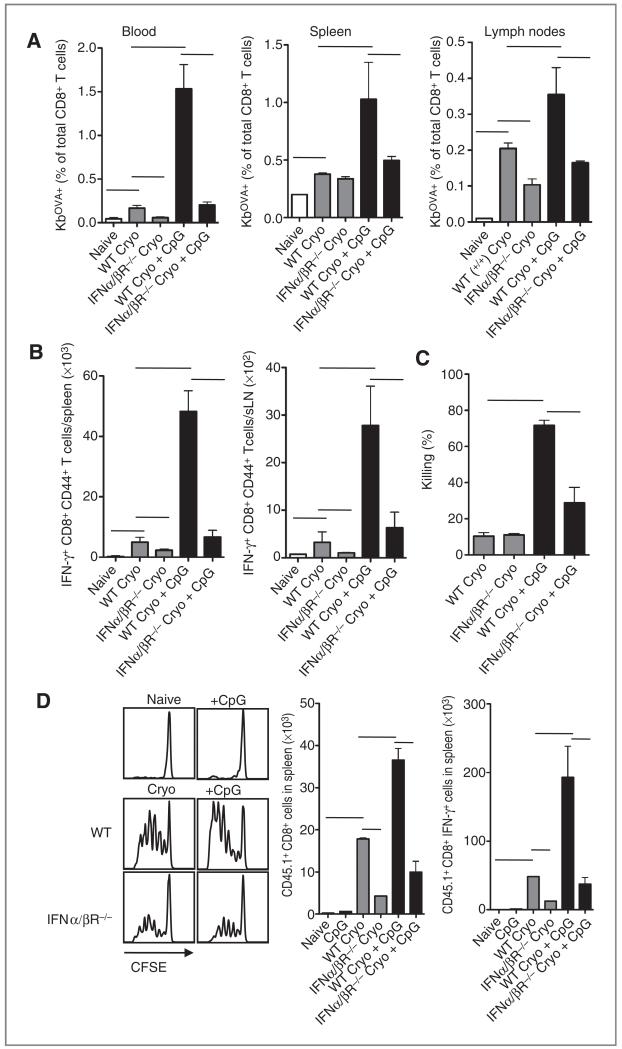
Figure 2.
Priming of endogenous CD8+ T cells and transferred OT-I T cells is abrogated in IFNα/βR−/− mice. A, B16OVA-bearing WT and IFNα/βR−/− mice were treated with cryoablation. After the ablation, indicated groups were additionally treated with CpG. Eight to 10 days after ablation, blood, spleen, and draining lymph nodes were isolated and stained with OVA-Kb tetramers. B, spleen and draining lymph nodes were restimulated with SIINFEKL peptide for 5 hours and analyzed for IFN-γ production. C, 6 to 8 days after ablation, mice received 5 × 10⁶ SIINFEKL-coated CFSE-labeled (low dose) splenocytes from C57Bl/6 SJL (CD45.1) mice and a similar number of CFSE-labeled (high dose) splenocytes coated with irrelevant peptide E1B192–200. After 18 hours, spleens were harvested and analyzed for the presence of CD45.1 CFSE-labeled cells. D, tumor-bearing mice were subjected to ablation with or without CpG and injected with CFSE-labeled purified CD8α+ OT-I SJL cells or WT cells as control population. Three or 4 days after transfer, CFSE dilution was analyzed in splenocytes. Histograms show representative data of cells gated on CD8, Vα2, and Vβ5. Bar graphs show mean values ± SEM of 4 to 6 mice and are representative of 2 similar experiments.

