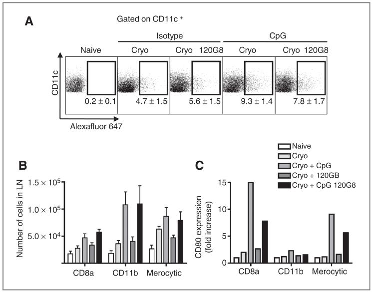
Figure 3.
Plasmacytoid DCs determine the function of cDCs. A, OVA-Alexa Fluor 647 (20 μg/20 μL) was injected into the tumor just prior to ablation with or without CpG. Two days after ablation, lymph node cells were analyzed for the uptake of Alexa Fluor 647 in CD11c+ cells. B, CD11c+ cells that were present in the draining lymph node were subfractionated into CD8α+ DCs (CD11c+B220−-CD24+CD172loCD11b−), CD11b+ DCs (CD11c+B220−CD24lo-CD8α−), and merocytic DCs (CD11c+B220−CD172loCD8α−-CD11blo). Bar graphs show mean values ± SEM of 4 to 6 mice. C, the relative increase in CD80 expression was determined in each subset.

