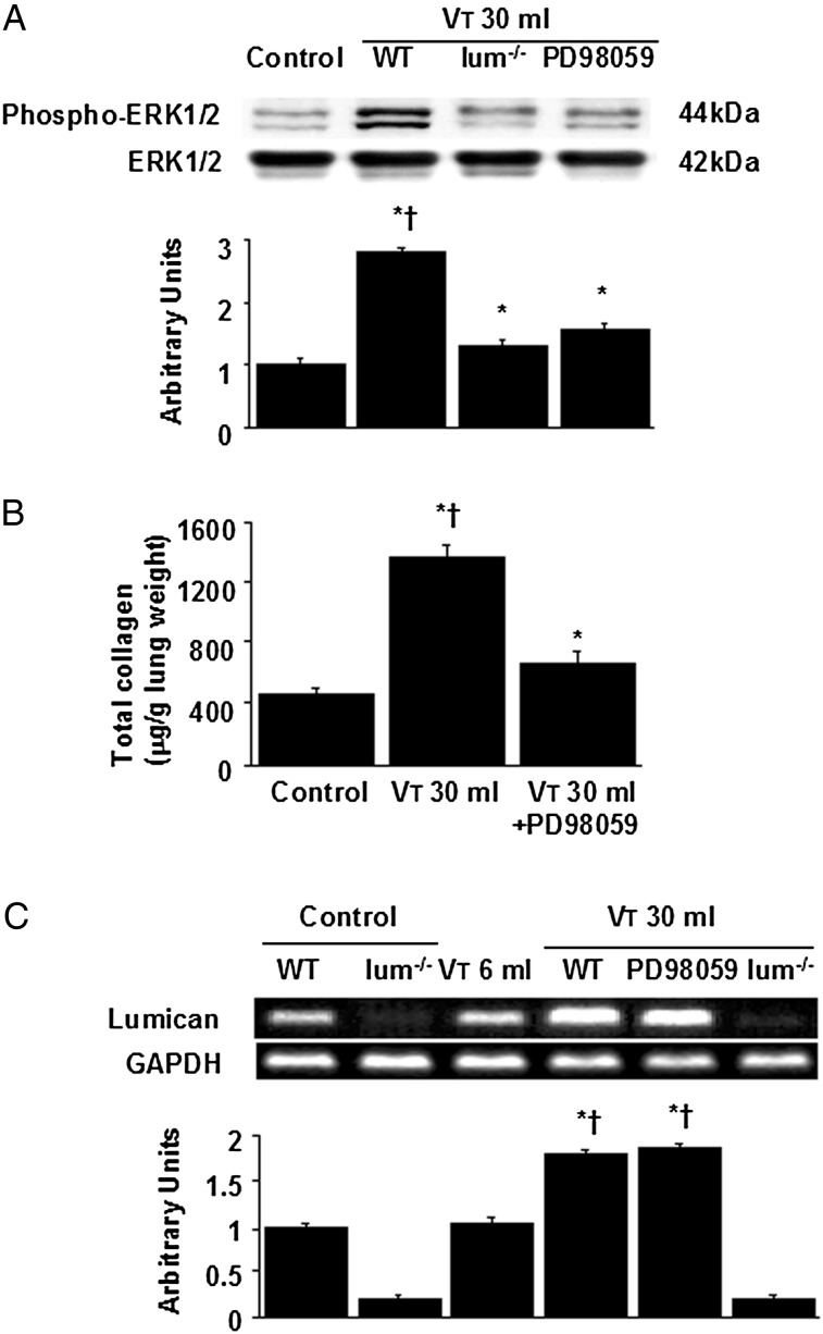
Figure 6.
Lum−/− mice and PD98059 reduced lung stretch-induced ERK1/2 activation, total collagen content, and lumican mRNA expression. A, Western blot was performed using an antibody that recognizes phosphorylated ERK1/2 and an antibody that recognizes total ERK1/2 expression in lung tissue from nonventilated control mice and mice ventilated at Vt 30 mL/kg for 2 h with room air. Arbitrary units were expressed as relative ERK1/2 phosphorylation (n = 5 per group). B, Total collagen level was from control mice and mice ventilated at Vt 30 mL/kg for 8 h with room air (n = 5 per group). P < .05 vs nonventilated control mice; †P < .05 vs PD98059 group. C, Reverse transcription-polymerase chain reaction assay was performed for lumican mRNA, GAPDH mRNA, and arbitrary units from nonventilated control mice and mice ventilated at Vt 6 mL/kg or Vt 30 mL/kg for 2 h with room air (n = 5 per group). Arbitrary units were expressed as the ratio of lumican mRNA to GAPDH. PD98059 2 mg/kg was given subcutaneously 30 min before ventilation. *P < .05 vs nonventilated control mice; †P < .05 vs lum−/− group. ERK = extracellular signal-regulated kinase. See Figure 1 legend for expansion of other abbreviations.

