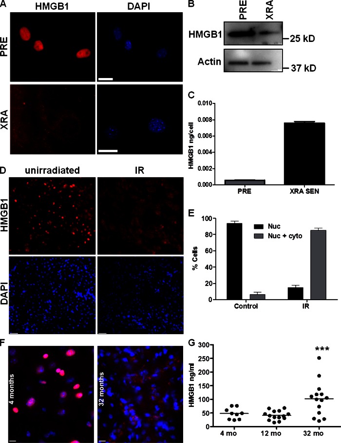
Figure 2.
HMGB1 relocalizes in SEN mouse cells. (A) PRE or SEN (XRA) MEFs were immunostained for HMGB1 (red). Nuclei were stained with DAPI (blue). Bar, 10 μM. (B) Lysates from PRE or SEN (XRA) MEFs were analyzed by Western blotting for HMGB1 and actin (control). (C) CM from PRE or XRA cells were analyzed for HMGB1 by ELISA. Error bars are the average of duplicate samples. (D) 6–8-wk-old C57BL/6 mice were unirradiated (control) or sublethally irradiated (IR). Kidneys collected 1 wk later were immunostained for HMGB1 (red); nuclei were stained with DAPI (blue). Bar, 25 μM. (E) Kidneys from mice in B were immunostained for nuclear (Nuc), or nuclear + cytoplasmic (Nuc + Cyto) HMGB1. More than 200 nuclei were scored blinded from 2–3 control or irradiated (IR) mice; error bars are SEM. (F) Kidneys from 4- and 32-mo-old C57BL/6 mice were immunostained for HMGB1 (red); nuclei were counterstained with DAPI (blue). Bars, 10 μM. (G) Sera from mice at the indicated ages were assayed for HMGB1 by ELISA. Kruskal-Wallis test was used to determine significance (***, P < 0.001).

