Figure 5.
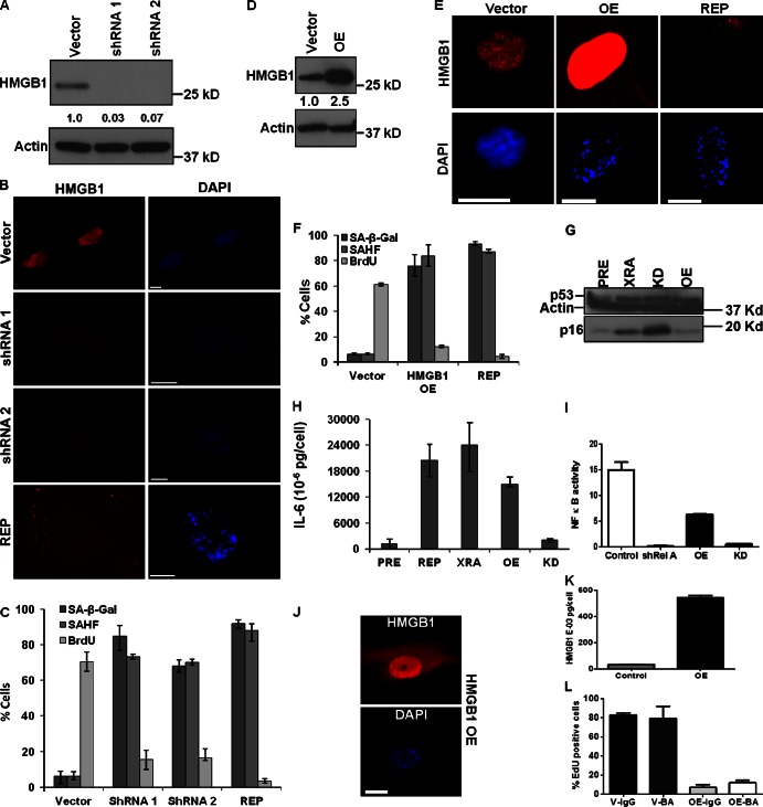
HMGB1 depletion or overexpression induces senescence. (A) Lysates from IMR-90 cells infected with lentiviruses carrying no insert (Vector) or HMGB1 shRNAs (shRNA1, 2) were analyzed for HMGB1 and actin (control) by Western blotting. Shown is expression level relative to Vector. (B) Cells in A were immunostained for HMGB1 (red). Nuclei were stained with DAPI (blue). REP SEN cells are shown for comparison. Bar, 10 µM. (C) Cells in B were scored for SA-β-Gal, BrdU (24 h pulse), and SAHF. Error bars = SEM of two independent experiments. (D) Lysates from IMR-90 cells infected with lentivirus expressing no insert (Vector) or HMGB1 (OE) were analyzed for HMGB1 and actin (control) by Western blotting. Shown is expression level relative to Vector. (E) Cells in D were immunostained for HMGB1 (red). Nuclei were stained with DAPI (blue). REP SEN cells are shown for comparison. Bar, 10 µM. (F) Cells in E were scored for SA-β-Gal, BrdU (24 h pulse), and SAHFs. REP SEN cells are shown for comparison. Error bars = SEM of two independent experiments. (G) Lysates from PRE IMR-90 cells or cells induced to senesce by XRA (XRA), HMGB1-overexpression (OE), or HMGB1-depletion (KD) were analyzed for p53, p16INK4a, and actin (control) by Western blotting. (H) CM from PRE or SEN (REP, XRA, and HMGB1 OE or KD) IMR-90 cells were analyzed for IL-6 by ELISA. Shown is a representative of two experiments. Error bars = SEM of duplicate determinations. (I) HCA2 cells expressing an NF-κB luciferase reporter were infected with lentiviruses carrying GFP shRNA (control), RelA shRNA, HMGB1 cDNA (OE), or HMGB1 shRNA (KD). 7 d after selection, shGFP and shRel A cells were given TNF for 24 h in serum-free media. Cells were lysed and luciferase activity measured (fold change over cells given BSA (control) protein). One-way ANOVA was used to analyze groups; P < 0.001. (J) Cells infected with HMGB1 OE lentivirus were stained for HMGB1 (red) and nuclei (DAPI; blue). Bar, 10 μM. (K) CM from cells infected with insertless (control) or HMGB1-expressing (OE) lentiviruses were assayed for HMGB1 by ELISA. Error bars = SEM of duplicate determinations. (L) IMR-90 cells infected with lentiviruses carrying no insert (V) or overexpressing HMGB1 (OE) were cultured for 5 d with mouse IgG or HMGB1 blocking antibody (BA) and assessed for EdU incorporation. Error bars = SEM of duplicate determinations.

