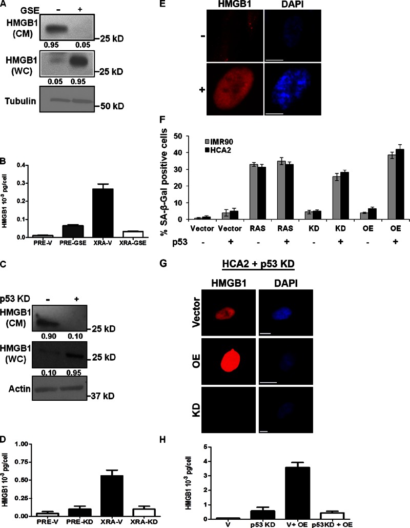
Figure 7.
HMGB1 relocalization and senescence caused by altered HMGB1 expression requires p53. (A) Lysates (WC) and CM from XRA SEN HCA2 cells infected with control (−) or GSE (+) expressing lentiviruses were analyzed for HMGB1 and tubulin (control) by Western blotting. The fraction of detectable protein is given below each lane. (B) CM from PRE or SEN (XRA) HCA2 cells expressing control (V) or GSE-expressing lentiviruses were analyzed for HMGB1 by ELISA. (C) Lysates (WC) or CM from XRA SEN IMR90 cells infected with control (−) or shp53 (+) expressing lentiviruses were analyzed for HMGB1 and actin (control) by Western blotting. The fraction of detectable protein is given below each lane. (D) CM from PRE or SEN (XRA) IMR90 cells expressing control (V) or shp53 (KD) lentiviruses were analyzed for HMGB1 by ELISA. (E) REP SEN HCA2 cells infected with control (GFP; −) or GSE (+) expressing lentivirus were immunostained for HMGB1 (red). Nuclei were stained with DAPI (blue). Bars, 10 µM. (F) IMR-90 and HCA2 cells expressing (+) or depleted of p53 (−) were infected with a lentivirus carrying no insert (Vector), oncogenic RAS, HMGB1 shRNA (KD), or HMGB1 (OE), and scored for SA-β-Gal 3 d later. Error bars = SEM of two independent experiments. (G) Cells in B were infected with lentiviruses carrying no insert (Vector), HMGB1 (OE), or HMGB1 shRNA (KD). 6 d after infection, cells were immunostained for HMGB1 (red). Nuclei were stained with DAPI (blue). Bars, 10 µM. (H) CM from cells infected with lentiviruses carrying no insert (V), shp53 (p53 KD), and/or HMGB1 (OE) were analyzed for HMGB1 by ELISA. Error bars = SEM of triplicate samples.

