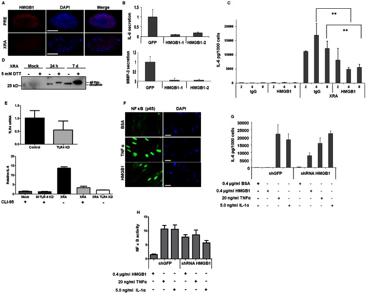
Figure 9.
HMGB1 regulates SASP factors. (A) PRE and SEN (XRA) WI-38 cells were immunostained for HMGB1 (red). DAPI stained the nuclei (blue). Images were uniformly enhanced to detect HMGB1 in SEN cells. Bar, 10 µM. (B) XRA SEN WI-38 cells were infected with lentiviruses carrying GFP or either of two HMGB1 (HMGB1-1, HMGB1-2) shRNAs. 4 d later, CM was collected and analyzed by ELISA for IL-6 and MMP-3. Shown are fold changes relative to shGFP. Error bars = SEM of two experiments. (C) PRE or SEN (XRA) HCA2 cells were incubated with 2, 4, and 8 µg/ml mouse IgG or HMGB1 blocking antibody. CM were analyzed by ELISA for IL-6. Error bars = SEM of triplicate samples. **, P < 0.02. (D) CM was collected from IMR90 cells that were mock irradiated, or 24 h or 7 d after irradiation. CM were heated in the presence (+) or absence (−) of 5 mM DTT, then analyzed by Western blotting. Positions of reduced (all thiol) and oxidized (disulfide) HMGB1 are indicated. (E) IMR90 cells were infected with control or shTLR-4 expressing lentiviruses and assessed for relative TLR-4 mRNA and IL-6 secretion. IL-6 secretion was assessed in mock (M) or X-irradiated (XRA) SEN cells expressing no insert or TLR-4 shRNA (TLR4 KD), or cultured in the presence (+) or absence (−) of CLI-95, a TLR-4 inhibitor. Shown is the fold expression relative to mock-irradiated cells without CLI-95. Error bars = SEM of triplicate samples. (F) IMR90 cells were cultured with 400 ng/ml BSA, 20 ng/ml TNF, or 400 ng/ml recombinant HMGB1 for 20 min and immunostained for NF-κB (p65 subunit; green). DAPI stained the nuclei (blue). Bar, 10 µM. (G) HCA2 cells infected with shGFP or shHMGB1-expressing lentiviruses were selected for 7 d and cultured for 24 h with indicated concentrations of BSA, recombinant HMGB1, TNF, or IL-1α. CM were analyzed by ELISA for IL-6. Error bars = SEM of triplicate samples. (H) HCA2 cells expressing an NF-κB luciferase reporter were infected with lentiviruses expressing shGFP or shHMGB1. 7 d after selection, cells were cultured for 24 h with the indicated proteins in serum free media at the concentrations in G. Cells were lysed and luciferase was measured. Luciferase levels are fold changes over cells incubated with BSA. One-Way ANOVA was used to analyze the groups; P < 0.001.

