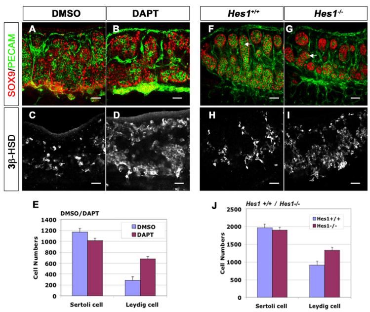
Fig. 2. Loss of Notch signaling results in an increase in fetal Leydig cells.
The coelomic domain of the gonad is upwards, anterior is leftwards and posterior is rightwards. (A–E) 11.5 dpc mouse XY gonads were cultured in DMEM for 48 hours with DMSO (A,C) or the γ-secretase inhibitor DAPT (B,D). (A,B) Sertoli cells were stained with antibodies against SOX9 (red), and vasculature and germ cells, with antibodies against PECAM1 (green). (C,D) Leydig cells, detected with antibodies against 3β-HSD were significantly increased (138%; n=8 gonads; P<0.001) in DAPT-treated gonads (D,E), compared with the DMSO control (C,E). Numbers of Sertoli cells are not significantly affected (E; n=3 gonads; P=0.1792). (F–J) Deletion of the Notch downstream target gene Hes1 (Hes1−/−) resulted in normal numbers of SOX9-positive Sertoli cells (red, G,J; n=3 gonads; P=0.2138), but an increase of 3β-HSD-positive Leydig cells (I,J; 47%; n=3 gonads; P=0.039) and a loss of germ cells (PECAM1, green; white arrows in F,G). Data presented are mean±s.e.m. of cell numbers from at least three independent gonad samples. Scale bars: 50 μm.

