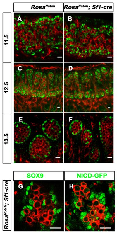
Fig. 3. Constitutively active Notch signaling in Sertoli cells does not change Sertoli cell fate.
The coelomic domain of the gonad is upwards, anterior is leftwards and posterior is rightwards.
(A–F) Immunofluorescent staining of PECAM1 (red) and SOX9 (green) on wild-type (A,C,E) and RosaNotch; Sf1-cre (B,D,F) mouse gonads at 11.5 (A,B), 12.5 (C,D) and 13.5 dpc (E,F). SOX9 appeared normal at all stages, although some mutant cords were smaller than wild type at 13.5 dpc (F). (G,H) Immunofluorescent staining of SOX9 (green, G) and GFP (green, a marker for NICD expression, H) on two adjacent sections of a 13.5 dpc mutant gonad showed that SOX9-positive Sertoli cells were also Notch active cells. Scale bars: 20 μm.

