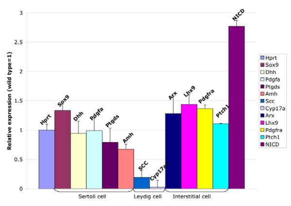
Fig. 6. Sertoli cell markers are not significantly different in RosaNotch; Sf1-cre gonads, but markers of differentiated Leydig cells are significantly downregulated.
mRNA was isolated from a single pair of mouse gonads, and expression was compared by Q-RTPCR between RosaNotch; Sf1-cre and wild-type gonads, normalized to Hprt. Sertoli cell-specific genes were similar (Sox9, Dhh, Pdgfa and Ptgds) or significantly downregulated (Amh) relative to normal expression levels (P=0.0534, 0.7942, 0.964, 0.4774 and 0.0323, respectively). Two differentiated Leydig cell genes, Scc and Cyp17a1, were significantly decreased in the mutant testis (P=0.0057, 0.00460). Interstitial markers, Arx, Pdgfra and Ptch1, were similar to wild type, while Lhx9 was significantly increased in RosaNotch; Sf1-cre gonads (P=0.0881, 0.2915, 0.5526 and 0.0212, respectively). NICD was significantly upregulated as expected. Data presented are mean±s.e.m. of Q-RT-PCR results from a total of four independent pairs of mutant and control gonad samples (each sample repeated at least three times for each gene).

