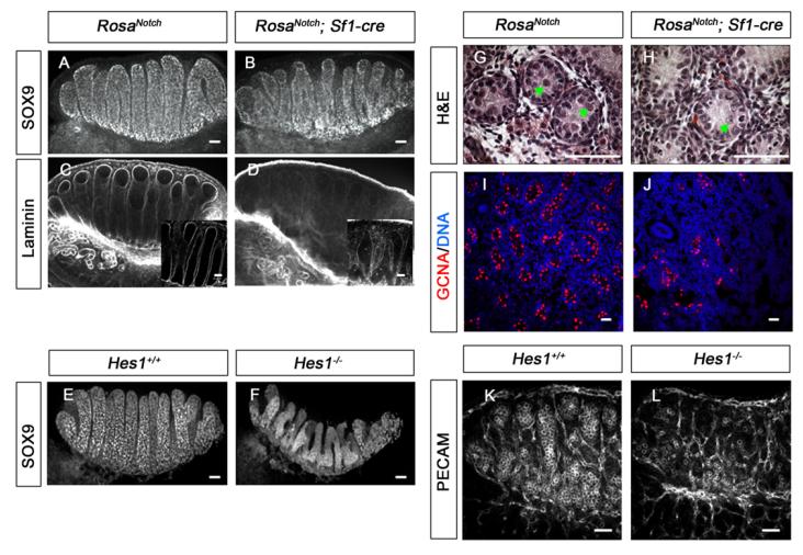
Fig. 7. Testis cord formation and germ cell survival are affected by gain or loss of Notch signaling in gonadal somatic cells.
The coelomic domain of the gonad is upwards, anterior is leftwards and posterior is rightwards. (A–F) Immunofluorescent staining for SOX9 (A,B,E,F) and Laminin (C,D, box inserts show 40× images of a confocal section) were used to evaluate mouse testis cord structure. At 13.5 dpc abnormal cord formation was detected in both, RosaNotch; Sf1-cre gonads (B,D) and Hes1−/− gonads (F). (G,H) At postnatal day (P) 1, fewer germ cells were detected inside testis cords based on Hematoxylin and Eosin staining (green arrows), although Sertoli cells appeared normal. (I,J) A significant decrease in the number of GCNA-positive cells (red; n=3, P<0.001) reflects germ cell loss in RosaNotch; Sf1-cre gonad at P1 (Syto13 stains DNA, blue). (K,L) C57BL/6 Hes1−/− gonads showed a significant germ cell loss at 13.5 dpc based on PECAM1 fluorescent immunostaining (P<0.001, n=3). Scale bars: 50 μm.

