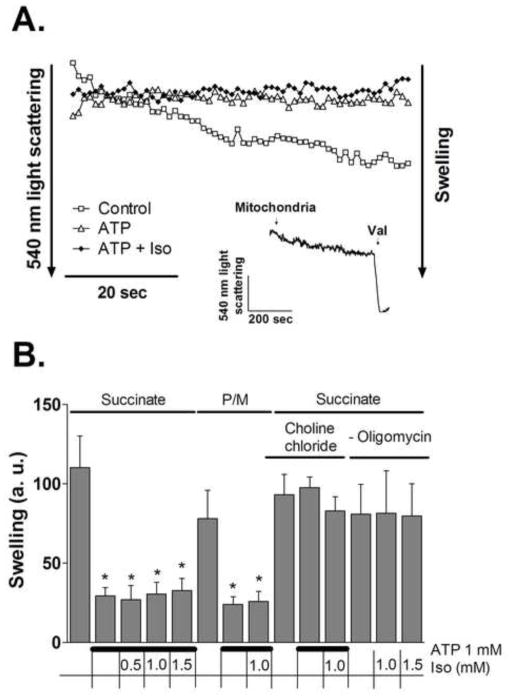
Figure 4.
Effect of isoflurane on mitochondrial swelling. (A) Representative light scattering traces of mitochondria (0.5 mg/ml) in K+ media supplied with 2 mM succinate. Where indicated, 1 mM ATP or 1 mM ATP plus 0.5 mM isoflurane (Iso) were present in the media from the beginning of the recording. Inset depicts maximum swelling induced by valinomycin (Val). (B) Magnitude of swelling, compared to control, determined as decrease in light scattering at 540 nm during 60 sec. Experimental conditions are listed below the graph. Values are means±S.D., n=4, *P<0.05 versus control.

