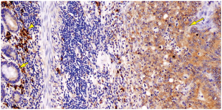
Figure 4.
Colon carcinoma cells (right, long arrow) show strong staining for GRP78 (also termed BiP), an ER chaperone induced by ER stress. Note the much weaker staining of surrounding normal enterocytes (left, short arrow). The intensely stained inflammatory cells in the background (arrowhead) are plasma cells.

