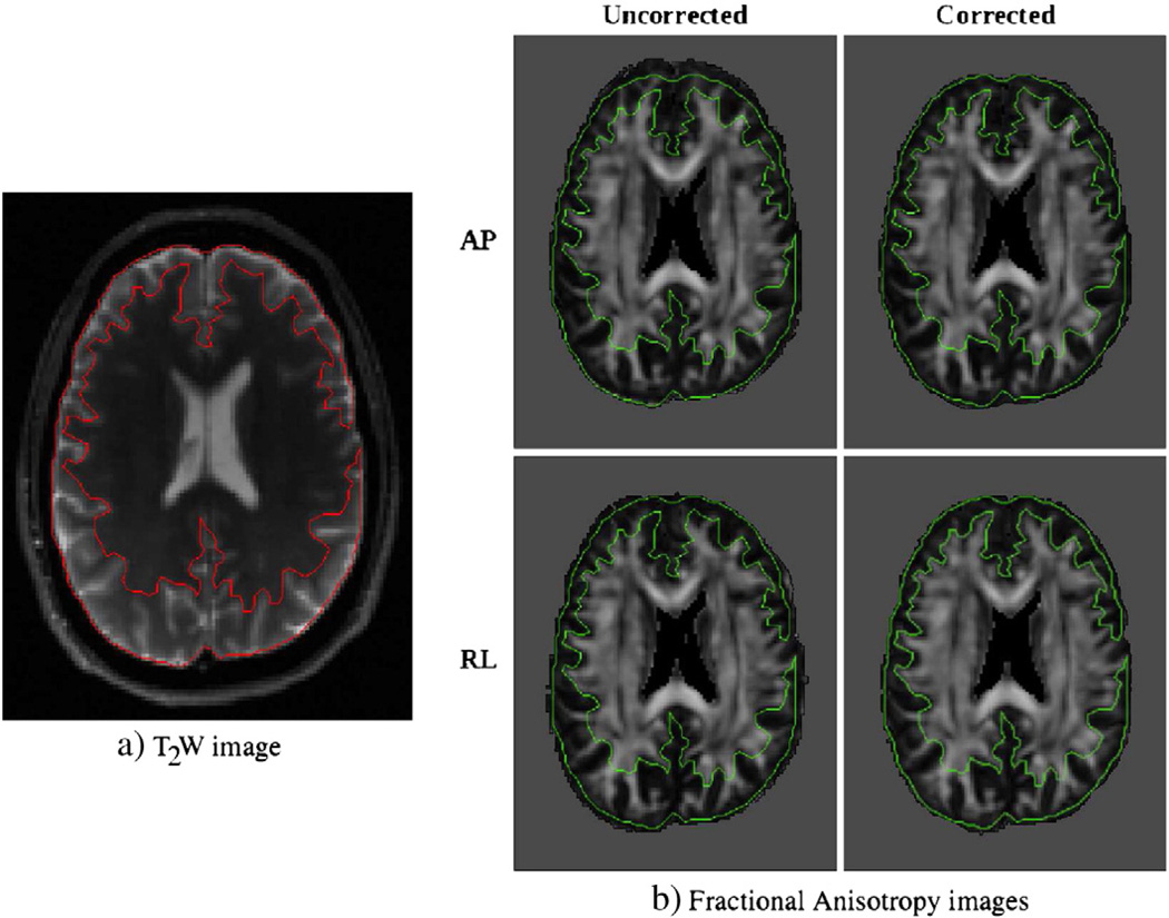
Fig. 1.
Outputs of registration for EPI distortion correction. The image on the left is the T2W structural image used as a target with boundaries indicated with a contour. Images on the right are the FA maps computed from data with different phase encode and correction schemes, with the same contour overlayed. For both uncorrected cases, the subject's brain extends out of the contour region in the direction of phase encoding, whereas this issue is minimized after elastic registration based correction.

