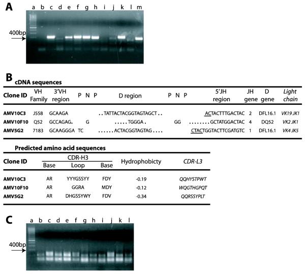
Figure 3. One step RT-PCR amplification of immunoglobulin gene transcripts from B cell cloning cultures.
Legend: (A) Samples containing B cells from cell cloning cultures, 0.66 cells/well, in the presence of S17 stromal cells (3000 cells/well), that scored positive for the secretion of IgM were subjected to one step RT-PCR for the amplification of VH transcripts using a promiscuous VH primer. The final RT-PCR products (~ 400 bp, lanes c-m) were identified by electrophoresis and are indicated in the figure with an arrow. In the gel shown, transcripts from 7 out of 11 IgM+ cultures were amplified (64 % efficiency): lane “a”, DNA ladder; lane “b”, culture with S17 alone. (B) mRNA from samples containing B cells from cloning cultures that scored positive for the recognition of the 190 kD antigen in mouse brain were subjected to one step RT-PCR for the amplification of transcripts of immunoglobulin V genes using a promiscuous primer as described. cDNA sequences: the VH family and the sequence of the heavy chain CDR3 (CDR-H3) are shown with the respective VL and JL genes for each clonotype. Clone ID, clone identifier. VH family, name according to the IMGT classification. 3′VH region, the most 3′ nucleotides of the VH (not including Cys = TGT). P-N-P, palindromic (P) and N region sequence. D region, DH gene sequence. 5′J-Region, the most 5′ nucleotides of the JH (not including Trp = TGG); underlined sequences can be from either of two germline genes. Dots represent nucleotides lost from the germline DH and JH gene segments. JH gene indicates the number of the JH element. DH gene, the DH element used; RF, the DH reading frame used. Light Chain, the Vκ Family and the Jκ usage of the light chain paired with the respective heavy chain is indicated. Predicted amino acid sequences derived from the cDNA sequences: Base, the predicted amino acid sequence of the CDR-H3 base. Loop, the predicted amino acid sequence of the CDR-H3 loop. Hydrophobicity, the average, normalized Kyte-Doolittle hydrophobicity of the CDR-H3 loop. CDR-L3, the predicted amino acid sequence of the light chain CDR-3. (C) A parallel set of cultures were done using rat thymocytes as feeder cells. Samples containing B cells from cell cloning cultures, 0.66 cells/well, that had scored positive for the secretion of IgM were submitted to one step RT-PCR as above. No RT-PCR amplification product could be detected (results representative of 3 different experiments).

