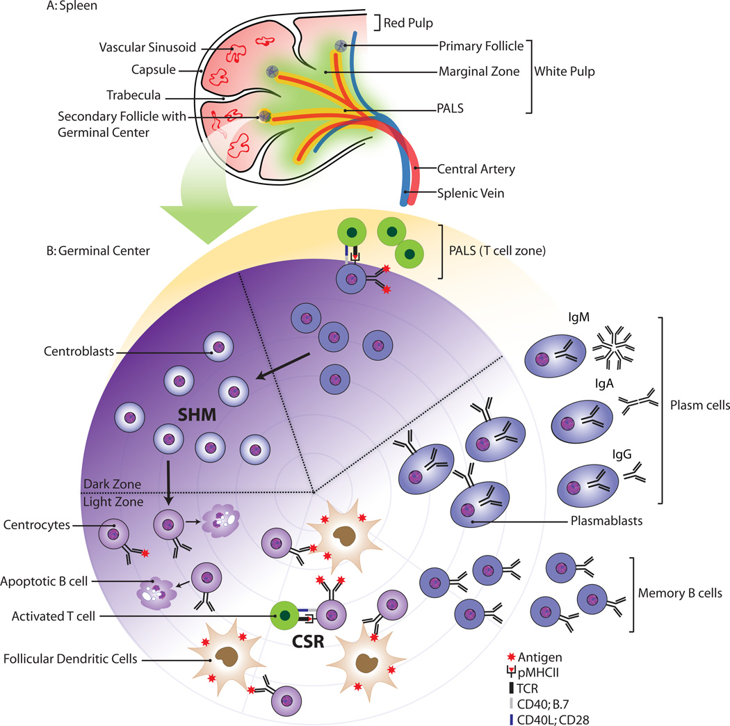
Figure 2.
Schematic morphology of the spleen (A) and the germinal center reaction (B). The white pulp consists of a central artery surrounded by T cells; the marginal zone and the follicles. Antigen-specific B cells and T cells interact at the border of the follicle. The B cells differentiate into centroblasts and undergo clonal expansion in the germinal center dark zone.

