
Figure 3.
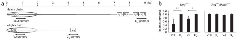
Uracil residues in VH and Vκ regions from mouse germinal center B cells. (a) Rearranged mouse Igh and Igk loci. Primers were designed to amplify 500-bp fragments spanning introns downsteam of V-gene segments rearranged to JH4 and Jκ2 gene segments, and genes encoding Cμ, and Cκ. Dotted ovals indicate the range of SHM on both loci. (b) Quantitative PCR analysis of intact DNA isolated from B220+GL7+ spleen cells of Ung−/− and Ung−/−Aicda−/− mice (immunized with KLH in adjuvant), then treated with UNG and APE1; results are presented as the difference in amplification of treated versus untreated DNA, relative to Gapdh amplification. *P = 0.008 and **P = 0.001 (two-tailed t-test). Data are from six experiments (error bars, s.e.m.).