Figure 1:

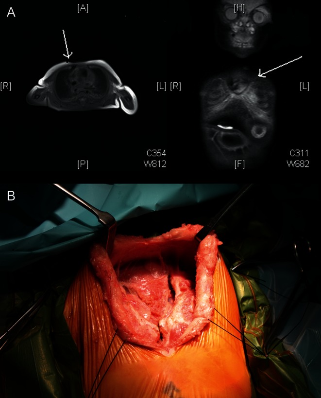
(A) Images of MRI showing the edges of lateral sternal bars (arrow). (B) Intraoperative photograph showing the lateral sternal bars and an inferior sternal attachment.

(A) Images of MRI showing the edges of lateral sternal bars (arrow). (B) Intraoperative photograph showing the lateral sternal bars and an inferior sternal attachment.