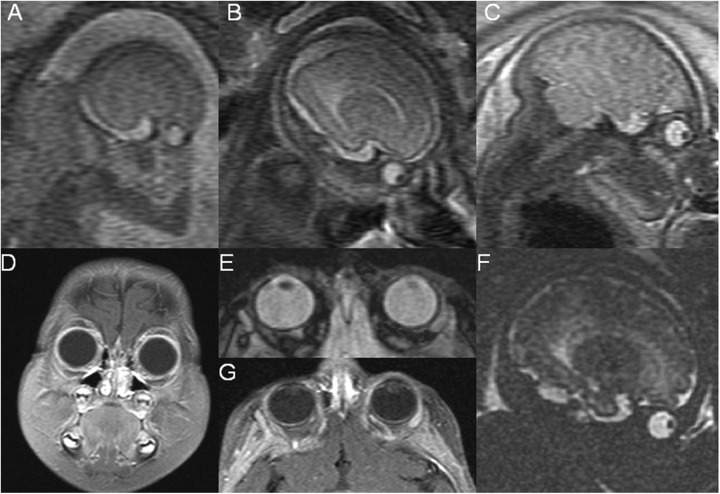
Figure 3.
Fetal and postnatal magnetic resonance (MR) images of patient 2 suspected by prenatal ultrasound to have an elevated intraocular mass at 37 weeks (Fig. 2) and confirmed on postnatal exam under anesthesia (Fig. 4). The sequential fetal MR imaging (A–C), single-shot fast-spin echo T2 sagittal views of fetal head and orbits performed at (A) 19 weeks, (B) 28 weeks, (C) 36 weeks show no evidence of an intraocular mass. A correlating hydrographic fetal MR sequence was performed at 36 weeks (G), which also confirmed no evidence of an intraocular mass. The postnatal MR imaging for this patient was performed 5 months after birth and after chemotherapy was given. No intraocular mass was detected on the postnatal MR exam (D–F), coronal fat saturated postcontrast T1 of the orbital (D), axial fat-saturated T2 imaging of the orbit (E), axial fat-saturated postcontrast T1 of the orbit (F).

