
Fig. 6.
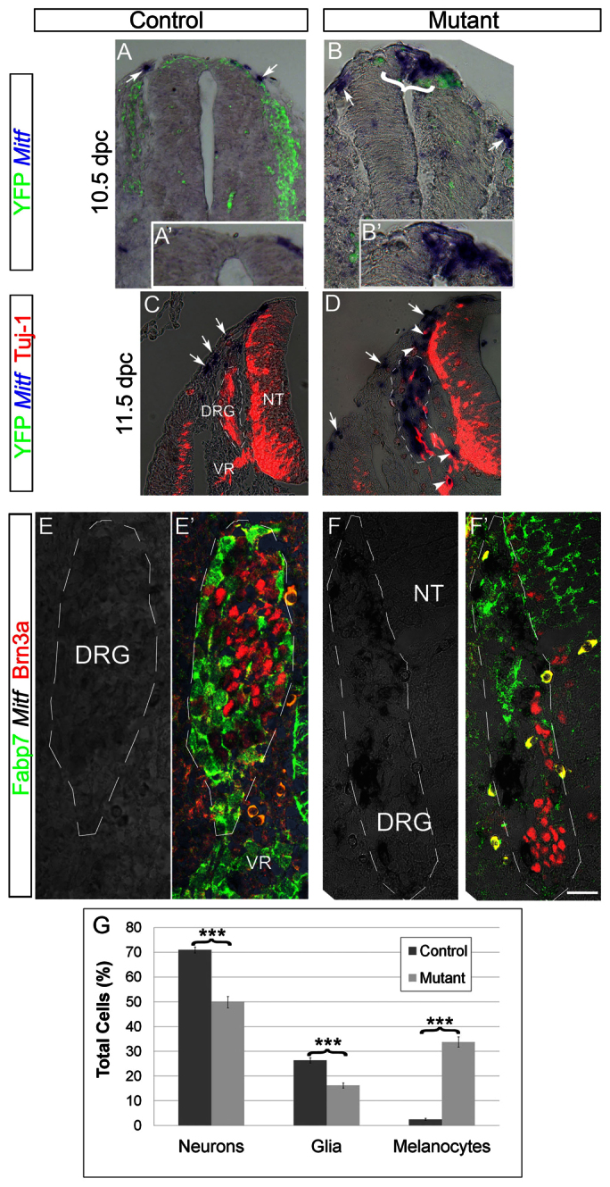
Ectopic expression of Mitf at the expense of neural traits in mutant mouse embryos lacking Foxd3 in the NC. (A-B′) Caudal to the hind limbs of 10.5 dpc controls, YFP+ NC migrate ventrally, and few Mitf+ cells are apparent in a dorsolateral position outside the NT (A,A′, arrows). Notably, in mutants, ectopic expression of Mitf is also seen in the dorsal NT (bracket in B, higher magnification in B′) in addition to emigrating NC (arrow in B). (C,D) In 11.5 dpc embryos Mitf+ melanocytes migrate ventral to the ectoderm in both controls and mutants (C,D, arrows). In controls, DRG cells express Tuj1 (C), whereas in mutants Mitf transcription is observed with a concomitant decrease in Tuj1 (D). Dorsal and ventral roots also contain Mitf+ cells in mutants (D, arrowheads). (E-F′) Expression of Mitf mRNA in mutant, but not control DRG, in comparison with staining for Brn3a and Fabp7. (G) Quantification of the percentage of melanocytes (Mitf+), neurons (Brn3a+) and glia (Fabp7+) of total labeled cells. Note melanocyte development at the expense of both neurons and glia (***P<0.005, n=40 and 37 sections counted in 12 ganglia of control and mutants, respectively). Scale bars: 35 μm in A,B; 20 μm in A′,B′; 130 μm in C; 100 μm in D; 30 μm in E-F′. VR, ventral root.