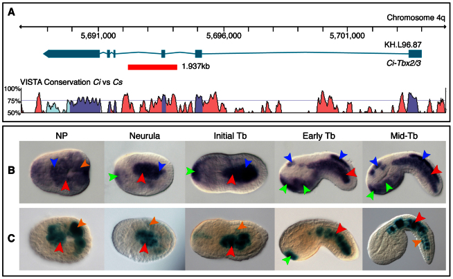
Fig. 2.
Identification of a CRM that recapitulates Ci-Tbx2/3 notochord expression. (A) Top: map of the Ci-Tbx2/3 locus. Exons and introns are denoted by teal rectangles and lines, respectively. A red box indicates the notochord CRM. Bottom: VISTA alignment illustrating the sequence conservation across the Ci-Tbx2/3 locus between Ciona intestinalis (Ci) and Ciona savignyi (Cs), obtained using the following parameters: calculation window, 100 bp; minimum conservation width, 100 bp; conservation identity, 50%. Purple peaks indicate conserved coding regions; light blue peaks indicate conserved 5′- or 3′-UTR; pink peaks indicate conserved non-coding regions. (B) Ci-Tbx2/3 whole-mount in situ hybridization. (C) X-Gal staining of C. intestinalis embryos carrying the Ci-Tbx2/3 notochord CRM in A. (B,C) Expression territories are highlighted with arrowheads: red, notochord; orange, muscle; blue, CNS; green, epidermis. In most panels, dorsal is upwards and anterior is towards the left. NP, neural plate; Tb, tailbud.

