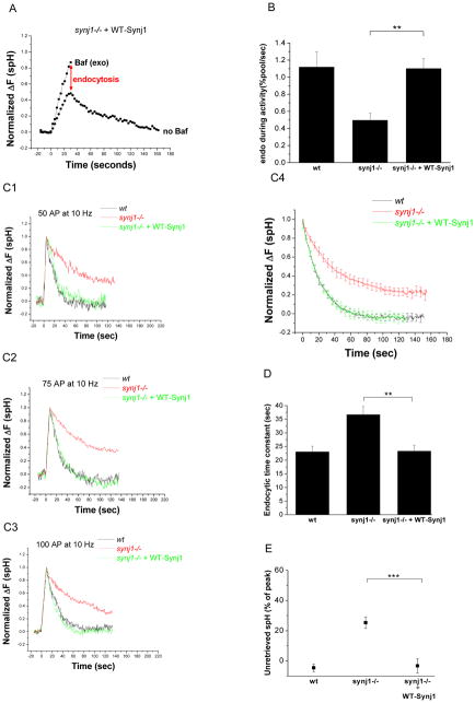
Figure 4. The endocytosis defects in synj1−/− neurons are corrected by the expression of wildtype Synj1.
(A) Synj1−/− neurons were co-transfected with spH and HA-tagged wildtype Synj1 (WT-Synj1). Representative example of the responses to a 10 Hz, 30 sec stimulus is shown here. The red arrow indicates the extent of endocytosis during persistent activity.
(B) The endocytic rate during 30 s activity in synj1−/− neurons transfected with WT-Synj1 is restored to wt levels. (n=8 for synj1−/− + WT-Synj1) **P<0.005, Error bars are SEM.
(C1–3) The endocytic time constant is restored by the expression of WT-Synj1. Representative examples of endocytosis after 50 AP (C1), 75 AP (C2), and 100 AP (C3) at 10 Hz in wt (black), synj1−/− (red), and synj1−/− + WT-Synj1 (green) conditions. Traces are normalized to the peak of the change in fluorescence.
(C4) The average endocytic time course for 50–100 AP in wt (black), synj1−/− (red), and synj1−/− + WT-Synj1 (green) conditions.
(D) The endocytic time constant for small stimuli is completely restored by the expression of WT-Synj1. **P<0.005
(E) The percentage of spH that remained unretrieved after 125s is restored to wt levels upon expression of WT-Synj1. ***P<0.0001
Averages from 19 wt, 30 synj1−/−, and 16 synj1−/− + WT-Synj1 expts. Error bars are SEM.

