Figure 1.
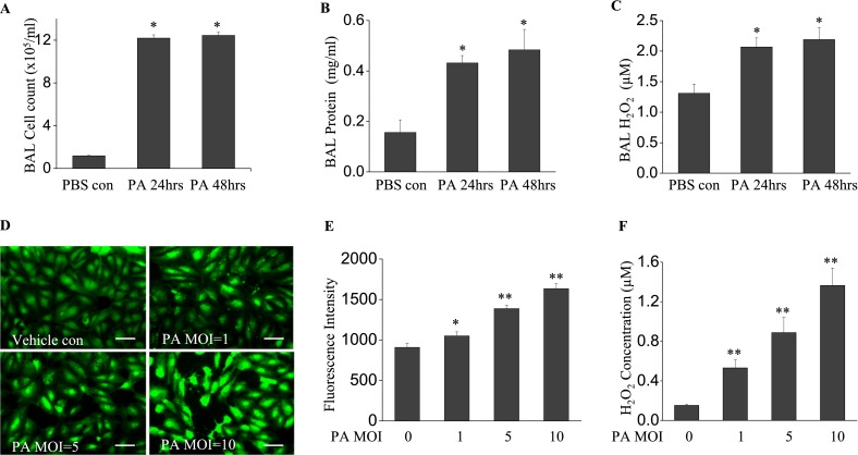
Pseudomonas aeruginosa (PA) induces lung inflammation and oxidative stress. Bronchoalveolar lavage (BAL) was collected at 24 and 48 hours after bacterial treatment. (A) A cell pellet was resuspended in 200 μl PBS for cell counting. (B) BAL protein and (C) H2O2 concentrations are shown. P. aeruginosa induced significant lung injury, as evidenced by increased BAL cell count, protein content, and reactive oxygen species (ROS) production. *P < 0.01, compared with PBS control samples (n = 4). (D) Human lung microvascular endothelial cells (HLMVECs), cultured in 35-mm dishes to approximately 90% confluence, were exposed to Pseudomonas aeruginosa strain 103 (PA103) at multiplicities of infections (MOIs) 1, 5, and 10 for 9 hours. Cells were then loaded with 5-(and-6)-carboxy-2',7'-dichlorodihydro-fluorescein diacetate (carboxy-H2DCFDA) (5 μM) for 30 minutes, and the oxidation of DCFDA was monitored under fluorescent microscopy (×200). Scale bars, 30 μm. (E) ROS production in each group was quantified. (F) H2O2 concentrations in culture media were assayed. PA103 induced significant ROS production in HLMVECs in a dose-dependent manner. *P < 0.05 and **P < 0.01, versus PBS control samples (n = 6). con, control.

