Figure 2.
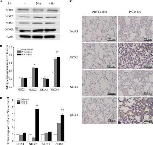
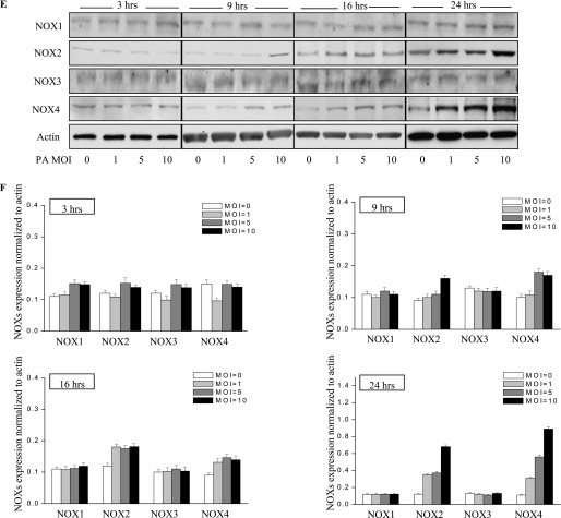
P. aeruginosa infection induces nicotinamide adenine dinucleotide phosphate–reduced oxidase–2 (NOX2) and NOX4 expression in murine lung tissue and HLMVECs. (A) Lungs were harvested at 24 hours and 48 hours for analyses of NOX proteins by Western blotting, or of mRNA by real-time PCR (D) (n = 4/group). Western blots were examined using an image analyzer, and NOX protein expression was quantified and normalized to total actin. (B) Approximately 2.5-fold and approximately 3.5-fold increases in NOX2 and NOX4 protein expression, respectively, were evident. *P < 0.01, compared with PBS control samples. #P < 0.01, compared with PBS control samples. (C) Immunohistochemical staining of NOX proteins. Scale bar, 200 μm. P. aeruginosa induced significant NOX2 and NOX4 protein expression in lung tissue, 48 hours after infection. However, it did not induce the expression of NOX1 or NOX3. (D) Real-time PCR quantification of NOX mRNA. **P < 0.01, compared with PBS control samples. ##P < 0.01, compared with PBS control samples. (E) HLMVECs were exposed to PA103 at MOI = 1, 5, and 10 for the indicated periods of time. Total cell lysates were subjected to SDS-PAGE and Western blotting for NOX protein expression. Actin was blotted to monitor protein loading. PA103 induced NOX2 expression in HLMVECs as early as 16 hours and 24 hours. MOI = 1 and 5 did not show significant NOX2 expression at 16 hours. PA also induced NOX4 expression at 16 hours and 24 hours after infection. However, PA103 did not induce NOX1 and NOX3 expression at any of the time points tested. The blot shown is representative of four independent experiments. (F) Statistical analysis for NOX proteins expression normalized to actin at various time points.


