Figure 4.
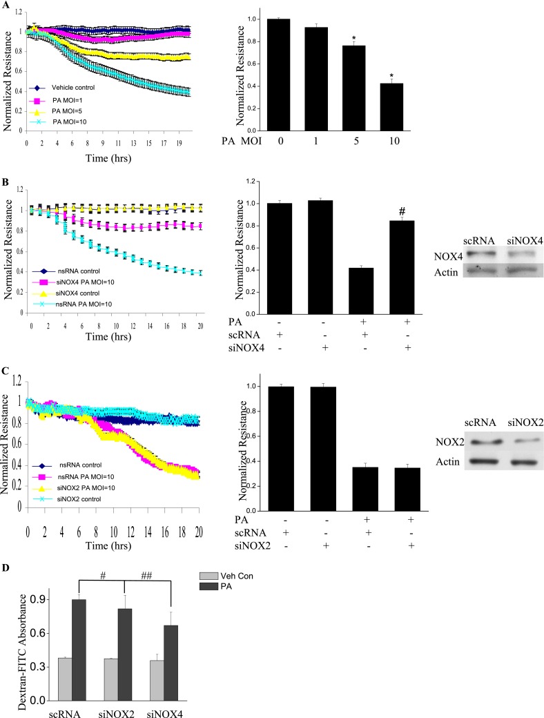
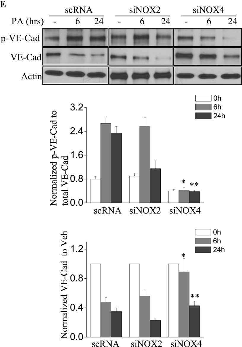
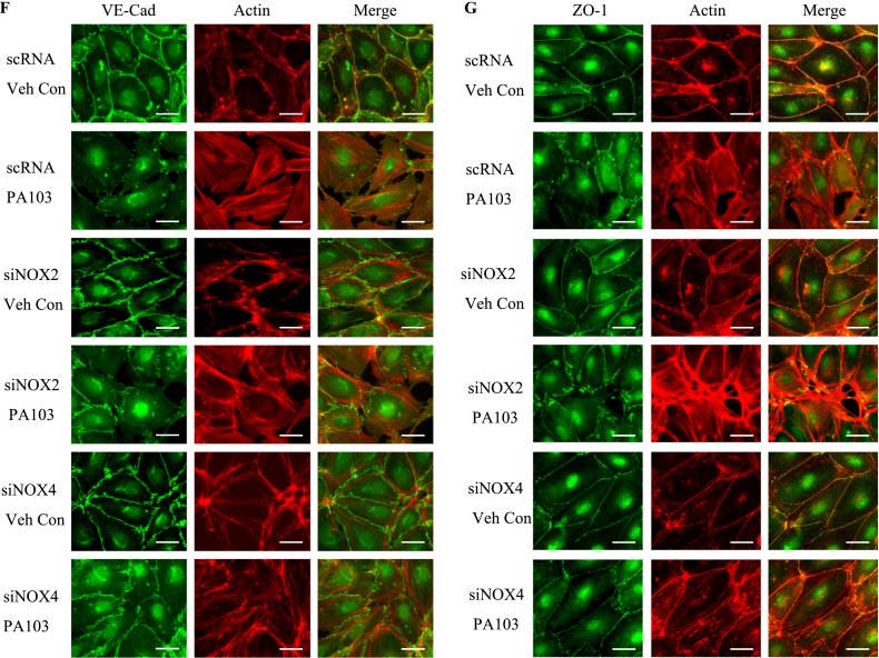
P. aeruginosa–induced barrier dysfunction in HLMVECs is blocked by NOX4 siRNA. HLMVECs were grown to confluence on gold electrodes and exposed to heat-killed PA103 at MOI = 1, 5, and 10. Transendothelial electrical resistance (TER) was monitored by electrical cell-substrate impedance sensor (ECIS). The basal monolayer TER was between 1,600 and 1,800 mΩ, and was normalized to 1 before initiating measurements. (A) P. aeruginosa significantly induced endothelial barrier permeability in a dose-dependent manner. *P < 0.01, versus PA MOI = 10. (B) HLMEVCs were transfected with scrambled siRNA or NOX4 siRNA (50 nM) for 72 hours before TER measurement. The P. aeruginosa–induced decrease in TER was partly blocked by NOX4 siRNA, compared with scrambled control siRNA–treated cells. **P < 0.01, versus scRNA PA MOI = 10. The knockdown of NOX4 was confirmed by Western blot, as shown below. (C) HLMEVCs were transfected with scrambled siRNA or NOX2 siRNA (50 nM) for 72 hours before TER measurements. The P. aeruginosa–induced decrease in TER was not blocked by NOX2 siRNA, compared with control siRNA–treated cells. The knockdown of NOX2 was confirmed by Western blotting, as shown below. (D) HLMVECs were first transfected with scrambled RNA, NOX2 siRNA, or NOX4 siRNA. Twenty-four hours later, cells were reseeded onto transwells, followed by P. aeruginosa challenge (MOI = 10). Permeability was indexed by the FITC-dextran leaked into the outside medium from the inner medium, 48 hours after reseeding. Only the knockdown of NOX4 inhibited the FITC-dextran leakage induced by P. aeruginosa. #P > 0.05, versus scRNA PA. ##P < 0.05, versus siNOX4. (E) NOX4 siRNA attenuated P. aeruginosa–induced VE-cadherin phosphorylation and decrease in expression, whereas NOX2 siRNA exerted no effect on VE-cadherin phosphorylation and expression. (F) Immunofluorescent staining of VE-cadherin, ZO-1, and actin was performed 24 hours after P. aeruginosa challenge (MOI = 10). For scRNA-transfected and siNOX2-transfected cells, P. aeruginosa induced significant stress fiber formation, and disruption of VE-Cadherin and ZO-1 from the cell adhesion junction, which was attenuated by NOX4 siRNA. Images are representative of the entire cell monolayer in two independent experiments. Scale bars, 10 μm. p-, phosphorylated; Veh Con, vehicle control.


