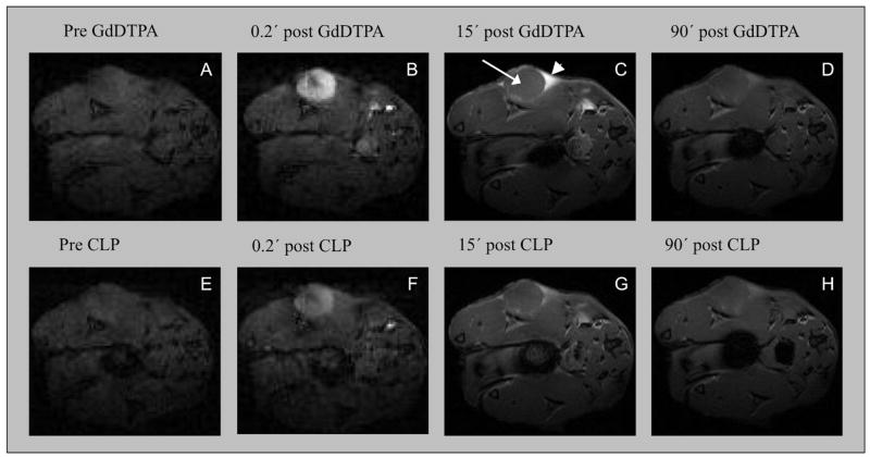
Figure 3.
Serial FLASH images pre- and post-injection of Gd-DTPA (top) and CLP (bottom) show transient tumor core enhancement following contrast administration, while regions of the tumor periphery show different enhancement kinetics. Arrow in image C indicates the tumor core and arrowhead indicates triangular enhanced region in tumor periphery. Washout of CLP from the periphery is 77% slower than Gd-DTPA due to its larger size, but both compounds show significantly faster washout than EP-2104R. Low resolution images A, B, E and F. High resolution images C, D, G and H.

