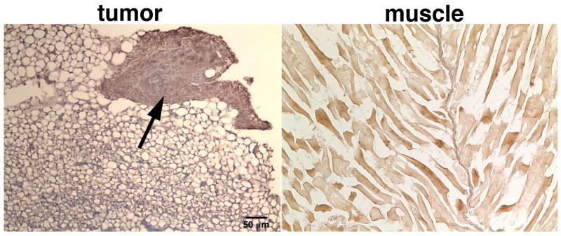
Figure 5.
Representative images of frozen tissue sections derived from the tumor (left) and adjacent muscle (right). The sections were stained with an HRP-conjugated fibrin-specific antibody, counterstained with DAB (brown) and visualized under light microscopy. Distinct fibrin-rich areas (arrow) could be identified at the periphery of the tumor. By contrast, DAB enhancement in the muscle tissue was at background levels.

