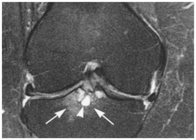
Figure 1.

Femoral notch bone marrow lesion (BML). Coronal proton density-weighted fat-suppressed magnetic resonance imaging (MRI) shows grade 3 WORMS (whole-organ MRI score) subchondral bone marrow lesion at the tibial subspinous region (arrows) with a cystic component (arrowhead). Image shows the BML at the tibial insertion of the anterior cruciate ligament. There is medial and lateral meniscal subluxation and partial maceration of the body of the medial meniscus. There is a diffuse thinning of the cartilage of the central medial femur.
