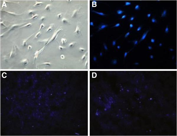
Figure 11.
DAPI positive cells were detected by fluorescent microscope. Compare with MSC infusion group, the US + Microbubble + MSC group has much more DAPI-positive cells localized in the ischemic myocardium (A-B). MSCs were labeled with DAPI, and all the cells were dyed in bright blue, and observed under light microscope (×200) and fluorescent microscope. (C) MSCs of US + Microbubble + MSC group (×100), (D) MSC infusion group (×100) [56].

