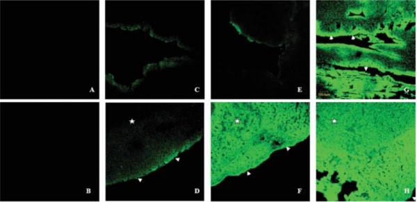
Figure 3.

The combination of UTMD and PEI in vivo. A, Phosphate-buffered saline group; B, phosphate-buffered saline + US; C, naked plasmid; D, plasmid + US; E, plasmid + liposome microbubbles (LM); F, plasmid + LM + US; G, plasmid + LM + PEI; H, plasmid + LM + PEI + US. There were no fluorescent signals in the negative control groups (A, B). When the mouse hearts exposed, the bases of the heart tissue samples had more transfected cells than the rest of the samples (D-H). Only a few fluorescent signals could be detected in the absence of US (E). A transmural fluorescent signal could be observed in the anterior wall after the microbubble was destroyed (F). EGFP was principally expressed in the subendocardial layer in the absence of US(C, E, and G and arrows in G). Combining with PEI and UTMD, the distribution of EGFP was not significant (H) [5].
