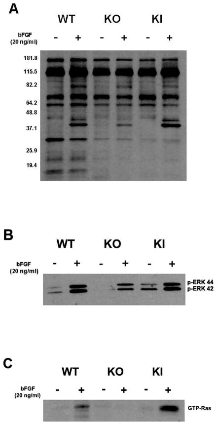
Figure 2.
Diminished bFGF-induced tyrosyl-phosphorylation substrates in KO cell lines. (A) WT, KO and KI cells were incubated in the presence or absence of bFGF for 5 minutes, and cell lysates were probed by Western blot for tyrosyl phosphorylation, and (B) in the presence of bFGF for 10 minutes before assessment of ERK phosphorylation. (C) Cell lysates from MT1-MMP WT, KO and KI fibroblasts were precipitated with GST-Raf beads, and samples were probed for GTP-Ras (after bFGF exposure for 30 minutes).

