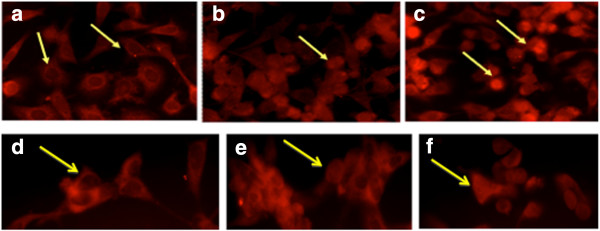
Figure 6.

NF-κB detection. Immunofluorescence of p65 NF-κB in PC3 (a, b, c) and LNCaP (d, e, f) cell lines. In untreated condition (a, d) NF-κB is 100% detected in the cytoplasm of cells (arrows). After LSESr (44 μg/ml) treatment (b, e) = 24 hours and (c, f) = 48 hours of incubation more than 30% of NF-κB translocated at nuclear level (arrows).
