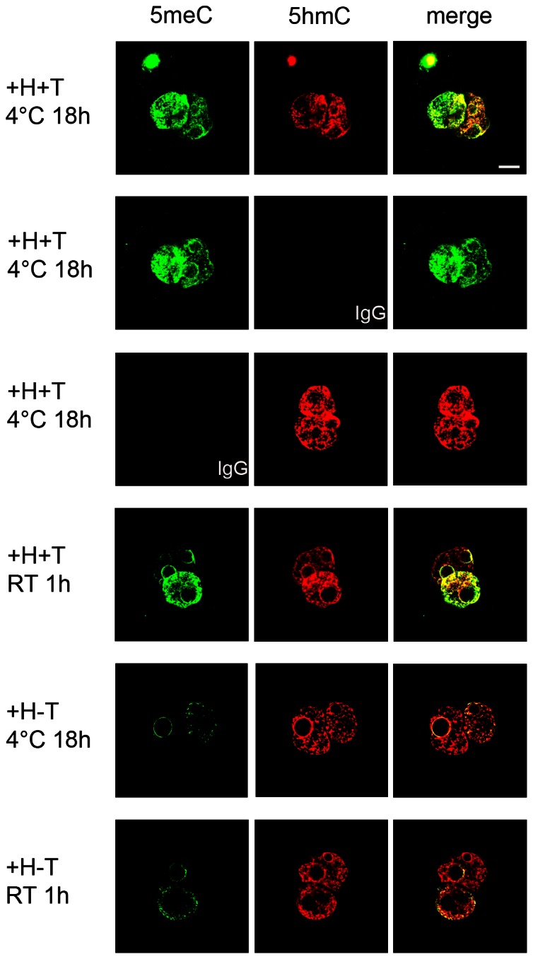
Figure 4. The effects of incubation time, temperature and epitope retrieval strategies on the simultaneous staining of 5meC and 5hmC.

PN5 stage zygotes were collected directly from the oviduct and fixed and prepared for staining. They were co-incubated with anti-5meC and anti-5hmC for 1 h at RT or 18 h at 4°C after epitope retrieval by either acid and trypsin treatment (+H+T) or acid treatment alone (+H-T). 5meC was detected with an FITC-labeled secondary antibody (green) and 5hmC by a Alexa Fluor633 (red) label. The images are representative of at least three independent replicates. The signals from the green and red channels were merged to detect co-localization. The bar is 5 µm and all images are at the same magnification.
