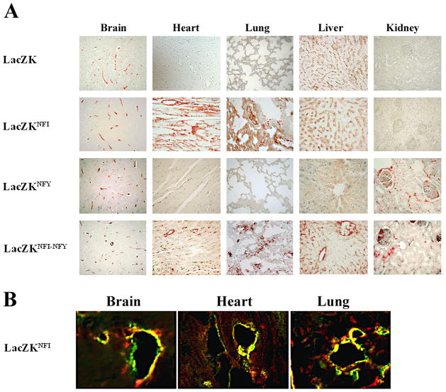
Figure 3. Immunohistochemical and immunofluorescence analyses of LacZ expression in organs of transgenic mice.
(A) Formalin-embedded sections (5 μm) of heart, kidney, liver, lung and brain from four transgenic mice (adult F1 generations) LacZK, LacZKNFI, LacZKNFY and LacZKNFI-NFY on tissue arrays were immunostained with anti-β-galactosidase antibody as previously described9 (magnifications 400X). Two littermates from each line and two independent lines for each transgene (except LacZK only one line) were analyzed and results from one representative line of each transgene shown. No staining was detected in sections from organs of non-transgenic mice (data not shown). (B) Sections (5 μm) of OCT-frozen brain, heart and lung from a LacZKNFI transgenic mice (line 92) were treated with anti-β-galactosidase (green) and anti-PECAM (red) antibodies to detect LacZ and endothelial cells as described (Materials and methods). The results are representative of two independent experiments using two littermates of this line (magnifications 400X).

