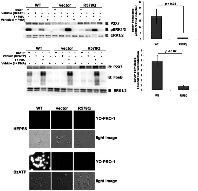
Figure 2. The P2X7 R578Q variant exhibits attenuated nucleotide-stimulated signaling and pore formation.
A and B, HEK293 cells expressing the P2X7 R578Q variant exhibit attenuated ERK1/2 activation. We have previously found that treatment with 1 µg/mL PMA plus 1 µg/mL ionomycin promotes ERK1/2 activation (data not shown), and this co-treatment was used here as a positive control. The error bars represent the standard error of the mean. These data are representative of at least three experiments. C and D, HEK293 cells expressing the P2X7 R578Q variant display decreased nucleotide-induced FosB/ΔFosB induction in comparison to wild-type P2X7. We have previously found that treatment with 1 µg/mL PMA plus 1 µg/mL ionomycin stimulates FosB/ΔFosB induction (data not shown), and this co-treatment was used here as a positive control. The top band on the FosB immunoblot is full-length FosB and the lower bands are its truncated splice variant ΔFosB [27]. The error bars represent the standard error of the mean. These data are representative of at least three experiments. E, The P2X7 R578Q variant displays drastically attenuated pore formation. These images are representative of at least three experiments. I = ionomycin.

