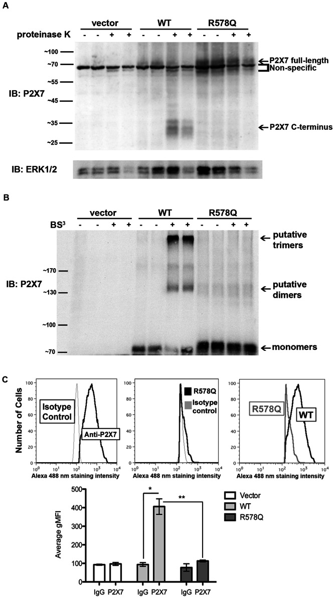
Figure 3. P2X7 R578Q is not expressed on the plasma membrane.
A, HEK293 stable cell lines were treated with the broad-spectrum protease proteinase K to detect the presence of the ∼30 kDa P2X7 C-terminal tail that is liberated when the extracellular domain of P2X7 is digested by protease when the receptor is localized at the plasma membrane. These data are representative of three experiments. B, HEK293 stable cell lines were treated with the cell-impermeable chemical cross-linker BS3 to detect P2X7 localization at the cell surface. The P2X7 monomers and putative dimers and trimers are indicated. The predicted molecular weight of human P2X7 is ∼69 kDa and it contains several N-linked glycosylation sites that result in slower mobility in SDS-PAGE gels [15]. Thus, P2X7 dimers are predicted to exhibit an apparent mass slightly greater than 140 kDa and P2X7 trimers are predicted to exhibit an apparent mass slightly greater than 210 kDa. These data are representative of at least four experiments. C, HEK293 cells were stained with an anti-P2X7 antibody or IgG isotype control, and the relative levels of surface-exposed P2X7 WT and R578Q were determined by flow cytometry. The graph represents the average of three independent experiments. * p<0.006, ** p<0.007. gMFI = geometric mean fluorescence intensity.

