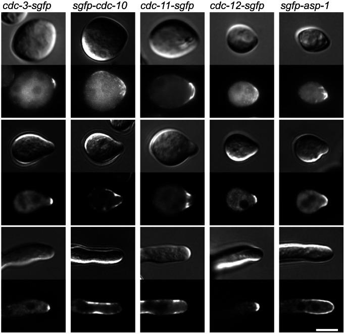
Figure 8. Septins show different patterns of localisation at germ tube tips.
Septin-GFP strains were incubated in liquid VMM for 3 h then imaged with DIC and widefield fluorescence microscopy to obtain representative images of germination and development. At the hyphal tip septins localised as a cap (CDC-3-GFP and CDC-12-GFP), an extended cap (GFP-ASP-1) or as a bar-like structures (GFP-CDC-10 and CDC-11-GFP). Scale bar, 5 µm.

