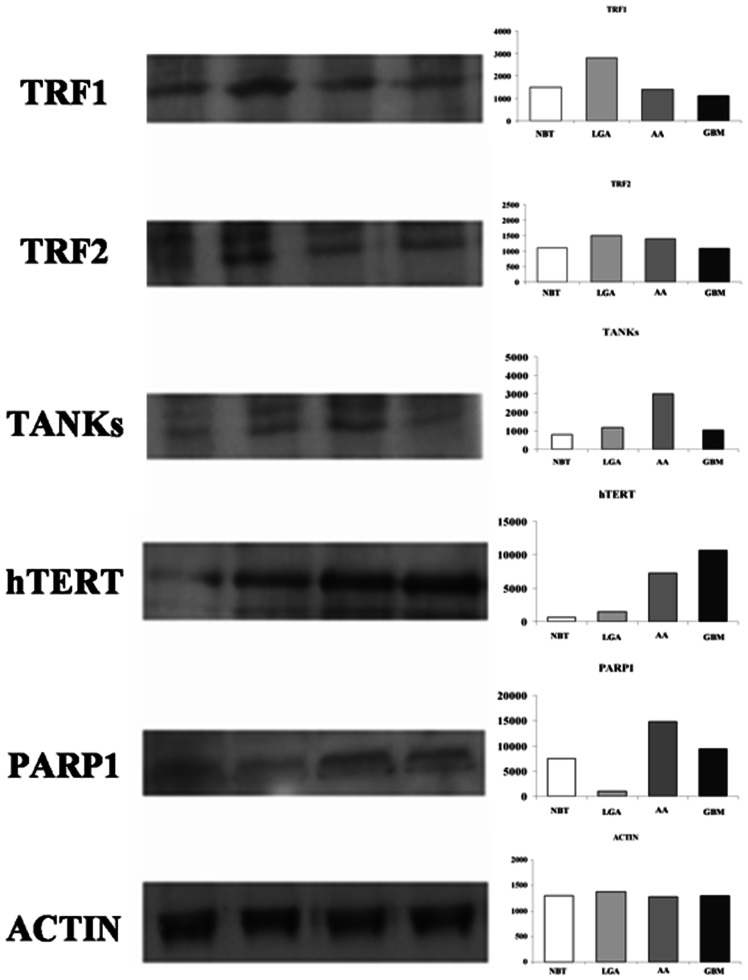
Figure 4. Western blot analysis of TRF1, TRF2, h-TERT, TANKs, and PARP-1.
Left: representative autoradiogram; right: graphs with quantitative data. Both TRF1 and TRF2 are over expressed in LGAs as compared with AAs and GBMs. h-TERT, TANKs, and PARP1 showed a tendency toward a higher expression in malignant astrocytomas (i.e. AAs and GBMs). Abbreviations: NBT: Normal Brain Tissue; LGA: Low grade Astrocytoma; AA: Anaplastic Astrocytoma; GBM: Glioblastoma Multiforme; TRF1, Telomeric repeat-binding factors 1; TRF2, Telomeric repeat-binding factors 2; h-TERT human telomerase reverse transcriptase; PARP1, Poly (ADP-ribose) polymerase 1.

