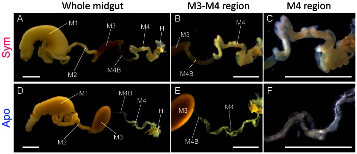
Figure 1. Dissected midgut of R. pedestris three days after fifth instar molt.
(A–C) Midgut of symbiotic insect. (D–E) Midgut of aposymbiotic insect. Abbreviations: M1, midgut first region; M2, midgut second region; M3, midgut third region; M4, midgut fourth region with crypts; M4B, anterior bulb of midgut fourth section; H, hindgut. Bars show 2 mm.

