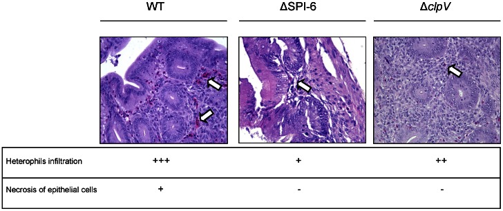
Figure 3. Histopathological changes in the cecum of infected chicks at day 3 post-infection.
Groups of 3 White Leghorn chicks were inoculated intragastrically by gavage with 109 CFU of the wild type S. Typhimurium 14028s strain, the ΔT6SSSPI-6 mutant strain or the ΔclpV mutant strain. At day 3 post-infection the chicks were sacrificed and the ceca were excised, fixed, stained with hematoxylin and eosin, and analyzed for histopathological lesions. Representative images of stained sections (400X) and scores for histopathological lesions in the cecum of infected chicks are shown (-, no changes; +, mild; ++, strong; +++, severe). White arrows indicate heterophil infiltration.

