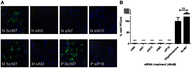
Figure 2. Reductions in HeV via pre-treatment with RNAi.
HeLa cells were transfected with 40 µg/ml Poly IC, 40 nM control or HeV targeting siRNAs. Transfection media was replaced after 4 hours and cells were infected with HeV clinical isolate at an MOI of 0.1. Infected cells were incubated for 24 hours before A) Infected cells were fixed and labelled for HeV N, P or M protein and DAPI stain. Cells were imaged with a Leica confocal microscope. Indicative images are shown. Scale bar = 50 µM. B) Supernatant was removed and titrated for TCID50 assay. Cells infected with supernatant were incubated for 3 days before TCID50 was calculated and shown as the mean ± S.E.M. of six biological replicates from two independent experiments. Significant differences between Oligofectamine control and siRNAs are indicated (***p = <0.001, two-sided t-test).

