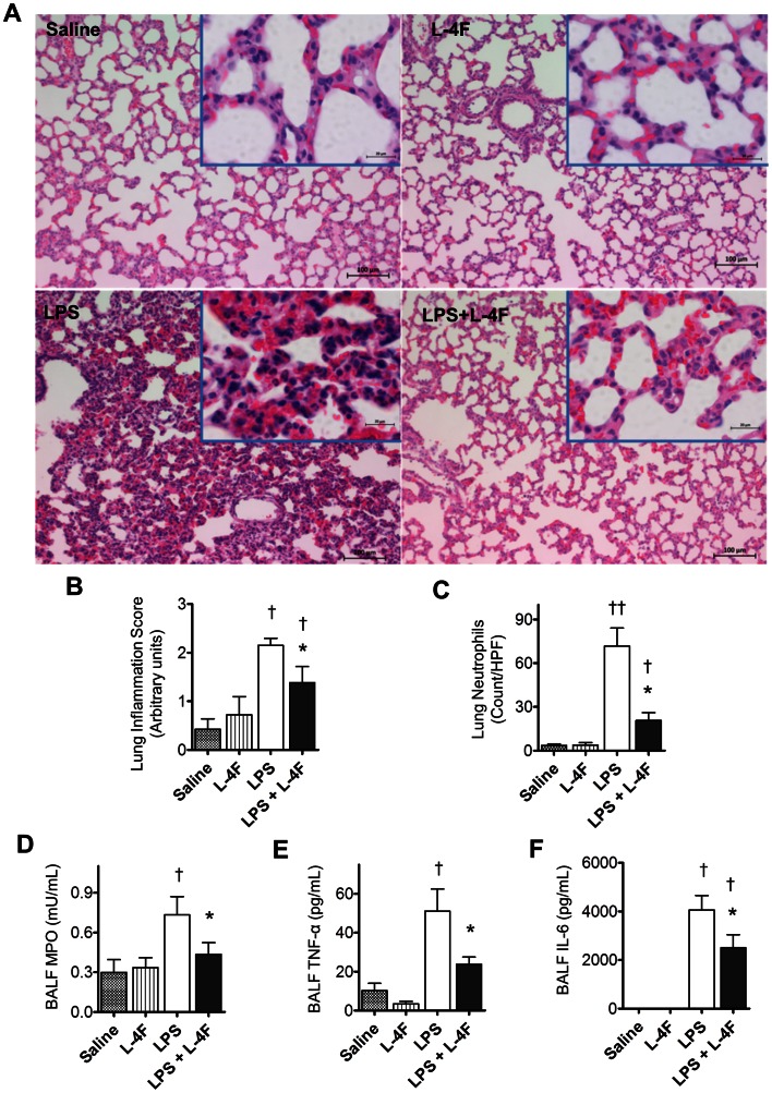
Figure 2. L-4F inhibits lung tissue inflammation in endotoxemic rats.
A: Lung H&E stained sections (magnification ×100, the insert is a magnification ×630). In saline treated rats, the alveolar walls appear normal and do not contain extensive numbers of polymorphonuclear leukocytes (PMNs). In L-4F treated rats, lung tissue is similar to control, but some PMNs are identified in alveolar capillaries In LPS treated rats, the alveolar walls are thickened and contain congested capillaries with numerous PMNs. Some of these cells have infiltrated outside the capillaries. In rats treated with L-4F, the lung is less affected than the lungs of rats receiving LPS and is similar to the lungs which received L-4F alone. Scale bars, 100 µm (in inserts, 20 µm). B: Blinded analysis of lung inflammation score in 5–8 rat/group. C: Neutrophil counts in lung parenchyma. The vast majority of the neutrophils were found in the septae, and fewer neutrophils were in the alveolar spaces. Neutrophil counts were performed based on the segmented morphology in high power fields (×40) in 30 measurements from 10 different randomly selected portal areas (n = 5/group). D, E, F: BALF MPO, TNF-α, and IL-6 respectively (n = 4 for controls, n = 8 for LPS groups). *P<0.05 vs. LPS, †P<0.05 and ††P<0.01 vs. Saline and L-4F.

